A little introduction
Protein Data Bank is a storage site for information and 3d images of biological proteins related macromolecule. It is worldwide, and shared among students and researchers, uploaded into the site and downloadable. One of the site is RCSB Protein Data Bank. The downloaded macromolecules images are opened by using a software like RasWIN, we could get the software from here.Samples of 5 proteins
LexA
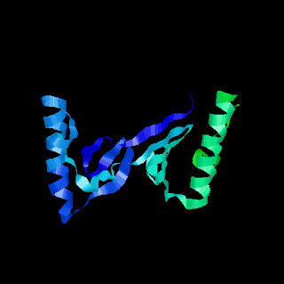
"Repressor LexA or LexA is a repressor enzyme (EC 3.4.21.88) that represses SOS response genes coding for DNA polymerases required for repairing DNA damage. LexA is intimately linked to RecA in the biochemical cycle of DNA damage and repair. RecA binds to DNA-bound LexA causing LexA to cleave itself in a process called autoproteolysis." - Wikipedia.orgLEXA G85D MUTANT
"LexA repressor undergoes a self-cleavage reaction. In vivo, this reaction requires an activated form of RecA, but it occurs spontaneously in vitro at high pH. Accordingly, LexA must ... " Click For More
| Type | Code |
| Molecule | LEXA REPRESSOR |
| Polymer | 1 | Type: Protein | Length: 202 |
| Chain | A, B |
| EC# | 3.4.21.88 |
| Mutation | G85D |
| Organism | Escheria coli |
| Gene Name | lexA exrA spr tsl umuA b4043 JW4003 |
| UnitProtKB | Protein Feature View | Search PDB | P0A7C2 |
For more information, visit http://www.rcsb.org/pdb/explore/explore.do?structureId=1JHF
Trypsin
"Trypsin (EC 3.4.21.4) is a serine protease found in the digestive system of many vertebrates, where it hydrolyses proteins. Trypsin is produced in the pancreas as the inactive proenzyme trypsinogen. Trypsin cleaves peptide chains mainly at the carboxyl side of the amino acids lysine or arginine, except when either is followed by proline. It is used for numerous biotechnological processes. The process is commonly referred to as trypsin proteolysis or trypsinisation, and proteins that have been digested/treated with trypsin are said to have been trypsinized." - Wikipedia.orgON THE DISORDERED ACTIVATION DOMAIN IN TRYPSINOGEN. CHEMICAL LABELLING AND LOW-TEMPERATURE CRYSTALLOGRAPHY

| Type | Code |
| Molecule | Trypsin |
| Polymer | 1 | Type: Protein | Length: 223 |
| Chain | A |
| EC# | 3.4.21.4 |
| Mutation | N/A |
| Organism | Bos Taurus |
| Gene Name | N/A |
| UnitProtKB | Protein Feature View | Search PDB | P00760 |
For more information, visit http://www.rcsb.org/pdb/explore/explore.do?structureId=2PTN
HtrA
Crystal Structure of DegP (HtrA)
"Molecular chaperones and proteases monitor the folded state of other proteins. In addition to recognizing non-native conformations, these quality control factors distinguish ... " Click for more
| Type | Code |
| Molecule | PROTEASE DO |
| Polymer | 1 | Type: Protein | Length: 448 |
| Chain | A, B |
| EC# | 3.4.21.107 |
| Mutation | S210A |
| Organism | Escherichia Coli |
| Gene Name | degP htrA ptd b0161 JW0157 |
| UnitProtKB | Protein Feature View | Search PDB | P0C0V0 |
For more information, visit http://www.rcsb.org/pdb/explore/explore.do?structureId=1KY9
Pepsin
"Pepsin (from the Greek πέψη, pepsi, meaning digestion) is an enzyme whose zymogen (pepsinogen) is released by the chief cells in the stomach and that degrades food proteins into peptides. It was discovered in 1836 by Theodor Schwann who also coined its name from the Greek word pepsis, meaning digestion (peptein: to digest). It was the first enzyme to be discovered, and, in 1929, it became one of the first enzymes to be crystallized, by John H. Northrop. Pepsin is a digestive protease, a member of the aspartate protease family." - WikipediaCRYSTAL STRUCTURE OF ASCARIS PEPSIN INHIBITOR-3
"The three-dimensional structures of pepsin inhibitor-3 (PI-3) from Ascaris suum and of the complex between PI-3 and porcine pepsin at 1. 75 A and 2.45 A resolution, respectively, have revealed ... " Click for more
| Type | Code |
| Molecule | MAJOR PEPSIN INHIBITOR PI-3 |
| Polymer | 1 | Type: Protein | Length: 149 |
| Chain | A |
| EC# | N/A |
| Mutation | N/A |
| Organism | Ascaris Suum |
| Gene Name | N/A |
| UnitProtKB | Protein Feature View | Search PDB | P19400 |
For more information, visit http://www.rcsb.org/pdb/explore/explore.do?structureId=1F32
Amylase
"Amylase /ˈæmɪleɪz/ is an enzyme that catalyses the breakdown of starch into sugars. Amylase is present in the saliva of humans and some other mammals, where it begins the chemical process of digestion. Foods that contain much starch but little sugar, such as rice and potato, taste slightly sweet as they are chewed because amylase turns some of their starch into sugar in the mouth. The pancreas also makes amylase (alpha amylase) to hydrolyse dietary starch into disaccharides and trisaccharides which are converted by other enzymes to glucose to supply the body with energy. Plants and some bacteria also produce amylase. As diastase, amylase was the first enzyme to be discovered and isolated (by Anselme Payen in 1833). Specific amylase proteins are designated by different Greek letters. All amylases are glycoside hydrolases and act on α-1,4-glycosidic bonds." - WikipediaHUMAN SALIVARY AMYLASE
"Salivary alpha-amylase, a major component of human saliva, plays a role in the initial digestion of starch and may be involved in the colonization of bacteria involved ..." Click for more
| Type | Code |
| Molecule | AMYLASE |
| Polymer | 1 | Type: Protein | Length: 496 |
| Chain | A |
| EC# | 3.2.1.1 |
| Mutation | 1 |
| Organism | Homo Sapiens |
| Gene Name | AMY1A AMY1 AMY1B AMY1C |
| UnitProtKB | Protein Feature View | Search PDB | P04745 |
or more information, visit http://www.rcsb.org/pdb/explore/explore.do?structureId=1SMD
No comments:
Post a Comment